Chain of Custody Form
Published: July 17th, 2009
Revised: February 13th, 2014
The Chain of Custody form ensures that every step with regards to our analysis process is reliable, confidential and secure.
Click here to download the form >
Fungal DNA barcode sequence identification [FDNA]
Published: July 8th, 2009
Revised: March 21st, 2023
This test provides a low-level identification of a pure fungal culture by sequencing of portions of the nuclear ribosomal gene, specifically the internal transcribed spacer (ITS) region or other gene regions deemed most suitable based on preliminary culture and microscopic screening. Although it is not always possible to recover fungi in pure culture in order to facilitate sequencing, where this can be accomplished, DNA-based sequence identification is the gold-standard for confirming microbial identification because it is capable of identifying rare or atypical fungal isolates irrespective of their responses to diagnostic culture media or biochemical tests.
DNA is isolated from cells using standard procedures. The nuclear ribosomal ITS is amplified by PCR and sequenced in both directions. A consensus sequence of both forward and reverse strands is constructed, and this sequence is used to query one or more fungal DNA sequence database tools. This approach to fungal identification is unmatched in its ability to accurately identify troublesome isolates, or to ascertain the taxonomic position of novel strains.
Laboratory code: FDNA
Service options

Blastomyces dermatitidis screen, bulk samples (qualitative) (PCR) [BLASTO]
Published: July 8th, 2009
Revised: April 19th, 2023
Blastomyces is a genus of dimorphic fungi that can cause a disease called blastomycosis in humans and other animals. Species of Blastomyces exist in the environment in a filamentous form and can produce infectious spores that can be inhaled. Once inside the body, the spores transform into yeast-like cells that can cause localized or disseminated infections. Blastomycosis primarily affects the lungs but can also involve other organs such as the skin, bones, and central nervous system. The disease is endemic in certain regions of North America, particularly in the Mississippi River and Great Lakes regions, and is more common in males than females. Two species are predominantly implicated as etiologic agents of this disease: B. dermatitidis and B. gilchristii. Both appear to be widespread throughout the endemic zone in North America.
This test expands on work conducted by Professor Tom Volk (now deceased) from the University of Wisconsin-Lacrosse. The procedure targets a portion of the virulence gene promotor, appropriately named BAD1 using conventional PCR. The results are reported as presence or absence of B. dermatitidis and B. gilchristii. A range of sample types can be analysed including dusts, bulk materials, and large volume air samples. In our experience, Blastomyces is extremely difficult to detect in environmental samples, and it seems likely that DNA of species of Blastomyces is highly ephemeral. As a consequence, this test is really only useful when an outbreak has been identified in humans or animals (especially dogs) as a means to assist in elucidating the nature of the exposure and reservoir. Even then, the timing of sampling must be as close as possible to the event in order to maximize the potential to yield informative positive results.
Laboratory code: BLASTO
Service options

Avian pathogen screen in air | bulk (Histoplasma, Cryptococcus & Chlamydophila by PCR) [APS]
Published: July 8th, 2009
Revised: March 21st, 2023
The fungi Histoplasma capsulatum and Cryptococcus neoformans are two agents of serious disease in otherwise healthy, immunologically normal humans and animals. Both agents are endemic in Canada, and both occur in the droppings of certain bird species in addition to other materials. Additionally, the bacterium Chlamydophila psittaci is the causative agent of psittacosis and is associated with bird feces (although in Canada this agent is predominantly restricted to captive parrots).
Histoplasma capsulatum is the causative agent of histoplasmosis, a non-communicable infectious disease that is mostly acquired by inhalation of environmental particulates. Greater than 95% of infections are clinically silent. These cases are detected sporadically by later serological tests or by the observation of small lung calcifications by x-ray. Clinically significant pulmonary histoplasmosis, in general, is often related to inhalation of a large inoculum load, or to the presence of an underlying immunodeficiency in the patient. Secondary blood-borne spread occurs in a small subset of cases, typically in people with serious pre-existing immune dysfunction. When treated, histoplasmosis generally responds well to systemic antifungal drugs. Histoplasma capsulatum occurs in accumulations of bat, pigeon or starling guano, particularly where those materials have come in contact with moist soil, where it grows as a cottony white mould until changing to a budding yeast-like phase in human or animal tissue. Only a few parts of Canada lie within the endemic distribution of H. capsulatum. Scattered cases and moderate to low levels of subclinical exposure are only found in southern areas of Quebec, Ontario, and Manitoba, as well as a few parts of Alberta, New Brunswick and Nova Scotia.
Members of the C. neoformans species complex are widely distributed basidiomycetous yeasts (more closely related to mushrooms than moulds) that occur in bat guano and bird droppings, particularly those of pigeons and chickens. Cryptococcus neoformans is an occasional cause of pneumonia and meningitis in people who inhale typically large quantities of infectious material. Immunocompromised people are more susceptible to cryptococcal disease, developing it occasionally following contact with birds and bird nesting materials.
Chlamydophila psittaci is a bacterium that can cause a respiratory disease in birds known as psittacosis or parrot fever. The bacterium can also infect humans who come into close contact with infected birds or their droppings. Outside of tropical regions, the agent is primarily restricted to captive birds especially parrots. Symptoms of psittacosis in humans include fever, headache, muscle aches, and a dry cough, which can progress to severe pneumonia if left untreated. Although rare, psittacosis can be a serious illness, especially for people with weakened immune systems.
The recognition and elimination of infective reservoirs of these agents is central to prevention. This test uses an extremely sensitive and specific PCR-based method to detect both agents in bulk material samples or large volume, time-integrated air samples collected on polycarbonate membrane filters. Bulk samples should be collected dry in a clean, new, food-grade, zippered freezer bag. Damp or moist samples should be refrigerated after collection prior to delivery to the laboratory. Air samples should be collected on a 25 or 37 mm 0.4 um membrane filter with a minimum volume of 1 cubic metre of air. The value of air sampling for these agents is likely to be low where there is little ongoing mechanical disturbance or aerosolization of dusts. In general, bulk samples are preferred for this test. Samples should be placed individually in a clean, new, food-grade, zippered freezer bag, and delivered to the laboratory within 24 hr of collection.
The detection of H. capsulatum is accomplished using primer sets that target portions of a catalase gene known as the “M-antigen”. Cryptococcus neoformans group is detected by primer sets that target a gene involved in capsular synthesis, CAP59. This method cannot distinguish between living and dead cellular material. Chlamydophila psittaci is detected by targeting a portion of the major outer membrane protein gene cluster (omp1). A negative result does not necessarily refute the presence of potentially infectious material in the area tested, since only a small amount of material is subjected to the test. The testing of multiple replicate samples is helpful to establish confidence on the generalizability of results. A positive result, however, may provide information that is useful in public health interventions.
More about histoplasmosis & crytococcosis >
Laboratory code: APS
Service options

Dust sample, quantitative fungal culture [FC-D]
Published: July 8th, 2009
Revised: March 24th, 2023
The analysis of vacuum collected dust is widely used to estimate microbiological exposures in human population health studies. In this context, dust analysis has repeatedly proven to be a useful proxy of exposure, because it serves as a reservoir for particles to which people are exposed in indoor environments. Dust itself also serves as a long-term integrative air sample, capturing transient airborne spore bursts, and retaining representative particles amid the growing bulk of textile fibres, hairs and skin scales that comprise the predominant mass-fraction of house dust.
Dust analysis is a useful adjunct to the investigation of indoor environments for fungal amplification sources. Although the total levels of culturable fungi in dust are not necessarily reflective of the condition of a building, careful evaluation of the species composition of the fungal cohort of dust can often lead a skilled investigator to meaningful revelations that otherwise would be missed. Although there are no guideline documents on the interpretation of dust sampling data, excellent baseline information for the expected fungal population of dust fungi is given in Dr. Scott’s PhD thesis, Studies on indoor fungi (2001).
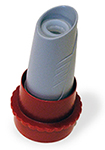
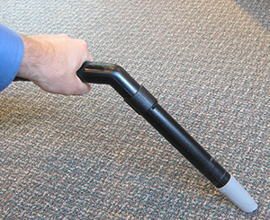
 Dust is usually obtained by vacuum collection from a predetermine area of flooring. Vacuum dust collectors range from very sophisticated devices, such as those used in research studies, to relatively simple “interceptor” filters that can be affixed to the sucking end of a household vacuum cleaner. The Dustream collector, pictured at right, is low-cost, widely accepted method for vacuum dust collection that does not require specialized equipment. The red adapter at the base allows to be attached to hoses of a range of sizes. This collection method can use used for culture-based tests, as well as endotoxin, glucan, and allergen assays such as mould and dust mite. Note that samples obtained from household vacuum cleaner bags are almost useless for biological analyses, since the typically growth-promotive environment within the collection bag, over time and depending upon the amount of moisture present and the frequency of cleaning and maintenance, causes changes in the composition and abundance of the captured fungal population.
Field collection method
- Don a pair of clean, disposable nitrile gloves. Use painter’s tape to outline a 1 m² area of flooring. If a contiguous space cannot be located to accommodate the full area, define as many sub-areas as necessary to demarcate a total floor area of 1 m².
 Remove the Dustream nylon mesh collection thimble from its polypropylene storage bag, and place the filter thimble into the collector shell. Affix the collector shell to the extension wand of the vacuum cleaner, using the red adapter ring, if necessary.
- Switch the vacuum cleaner on, and proceed to vacuum the entire surface within the demarcated area. To ensure thorough coverage, make 7 repeated passes over each stroke prior to proceeding to the adjacent stroke. Covering the entire area in this manner should take approximately 10 min. In the event that a sufficiently large amount of dust is present that the vacuum stalls or overheats due to back-pressure, record the actual area vacuumed to the nearest stroke in which 7 passes were completed.
- Remove the nylon mesh collection thimble from the collector shell, and place the thimble into a clean vial or polypropylene bag. Be sure to label the container with the sample number, job number, date and time of collection, and the surface area obtained. Use an indelible marker to ensure that the label data is not easily wiped off of the container. Wipe any residual fine dust from the collector shell using a 70% isopropyl alcohol wipe prior to beginning another sample collection.
Laboratory code: FC-D
Service options

Results reporting
The results of this test will be reported quantitatively, as Colony Forming Units per gram of dust (CFU/g), and broken down by species, species-group, or genus, depending on the taxon observed. For clients desiring to know the mass of total and/ or fine (< 105 um) dust recovered, we would be happy to supply these data for a small additional fee.


